

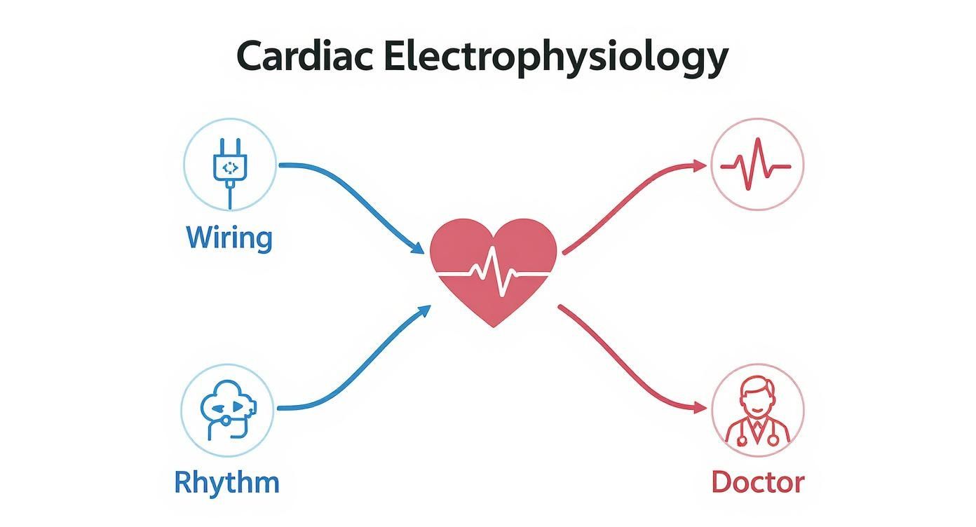
So, what exactly is cardiac electrophysiology? In simple terms, it’s a super-specialized field of cardiology that focuses entirely on your heart’s electrical system and the rhythm problems—called arrhythmias—that can pop up.
Think of it like this: your heart has its own internal wiring. When everything is working right, you don’t even notice it. But when there’s a glitch, you need a specialist. A cardiac electrophysiologist is basically a master electrician for your heart.
Your Heart’s Built-In Electrical System
At its heart (pun intended), your heart is an amazing pump, but it can only do its job because of a sophisticated electrical system that keeps everything in perfect time. Tiny electrical signals travel along specific pathways, telling your heart’s four chambers exactly when to squeeze and when to relax. This perfectly coordinated sequence is what we call a heartbeat.
It’s a bit like the wiring in your house. When the circuits are working properly, the lights stay on and your appliances run without a hitch. But if you have a short circuit or a faulty connection, you might get flickering lights or a total blackout. Your heart’s electrical system isn’t much different. A disruption can make it beat too fast, too slow, or just plain erratically. That’s an arrhythmia.
The Electrophysiologist: Your Heart Rhythm Specialist
A cardiac electrophysiologist, or EP for short, is a cardiologist who has gone through years of extra, intensive training focused just on heart rhythm disorders. They’re the detectives who figure out why your heartbeat is off.
While a general cardiologist looks at the big picture of your heart’s health—like blocked arteries or valve issues—an EP’s world is the electrical wiring. Their job boils down to three key things:
- Pinpointing the exact source of an abnormal heart rhythm.
- Figuring out your risk for more serious problems linked to that arrhythmia.
- Creating a personalized treatment plan to get your heart back into a healthy rhythm.
This razor-sharp focus is what allows them to use incredibly advanced tools and perform delicate procedures to fix those electrical short circuits, tackling everything from annoying palpitations to life-threatening rhythm disturbances.
An EP’s goal is to map out the heart’s electrical pathways, find the precise spot where the “glitch” is happening, and correct it. This restores stability and predictability to your heartbeat, which is crucial for both your safety and your quality of life.
Where Did This All Come From?
Studying the heart’s electricity isn’t a new idea. The field has a long, fascinating history, but it really took off back in 1902. That’s when a Dutch physician named Willem Einthoven invented the first practical electrocardiogram, or ECG.
For the first time, doctors could actually see the heart’s electrical activity on paper without having to do anything invasive. This machine gave us the classic P, Q, R, S, and T waves we still use today to represent a single heartbeat. Einthoven’s invention was the bedrock of modern electrophysiology. You can learn more about the history of clinical electrophysiology and its major turning points.
Today, EPs have taken that foundation and run with it. We now use technology that creates stunningly detailed, 3D maps of the heart’s electrical circuits in real-time. This level of precision allows us to deliver targeted treatments that were pure science fiction just a few decades ago, often permanently fixing rhythm problems and letting people get back to living full, active lives.
To really understand what is cardiac electrophysiology, you have to first appreciate the beautiful rhythm it’s all about. Every single heartbeat you feel is the end result of a perfectly timed electrical signal, a journey that starts and finishes in a fraction of a second.
Think of it like a lightning-fast relay race happening inside your chest, one that ensures your heart pumps blood exactly when and how it should.
The Starting Block: The SA Node
This race always kicks off from the same spot: the sinoatrial (SA) node. Tucked away in the upper right chamber of your heart (the right atrium), the SA node is your body’s own natural pacemaker. It creates a tiny electrical spark that sets the pace for the entire heart. This is the starting gun for the heartbeat.
Once that spark fires, the electrical signal ripples across the heart’s two upper chambers, the atria. This wave of electricity causes them to contract, squeezing blood down into the ventricles, the heart’s powerful lower chambers. It’s a smooth handoff, but the signal needs to take a quick, calculated pause before moving on.
The Gatekeeper: The AV Node
That strategic pause happens at the atrioventricular (AV) node, which acts as a crucial gatekeeper. Positioned right between the atria and the ventricles, the AV node holds onto the signal for just a moment.
Why the delay? It’s absolutely critical. This brief pause gives the atria enough time to finish their contraction and completely fill the ventricles with blood. Without this checkpoint, the chambers would contract on top of each other, and the pump would fail. The AV node makes sure the atrial and ventricular contractions happen in a perfect one-two sequence.
Once the ventricles are full and ready, the AV node releases the signal to continue the race.
The Final Sprint: Ventricular Contraction
From the AV node, the signal shoots down a specialized pathway called the Bundle of His, which immediately splits into right and left bundle branches. Think of these as highways carrying the impulse toward the bottom of the heart.
From there, the signal spreads like wildfire through a vast network of tiny Purkinje fibers embedded all through the ventricular muscle. This is the final, incredibly fast leg of the journey. The Purkinje fibers deliver the electrical message to every part of the ventricles at virtually the same time.
This synchronized jolt triggers a powerful, unified squeeze from the ventricles, which pumps oxygen-rich blood out to your brain and the rest of your body. And just like that, the heartbeat is complete.
The infographic below helps visualize these core components—the heart’s wiring, its rhythm, and the specialists who treat them.
After this powerful contraction, the heart muscle relaxes and the whole system instantly resets, ready for the SA node to fire the next impulse and start the cycle all over again.
The entire sequence—from the SA node’s initial spark to the final ventricular squeeze—is what we call a normal sinus rhythm. Every single component has a vital job. If there’s a problem at any point in this pathway, the heart’s rhythm can be disrupted, leading to what’s known as an arrhythmia.
Knowing how this perfect sequence is supposed to work is the first step in figuring out what’s going wrong. When an electrophysiologist looks for a rhythm problem, they’re essentially a detective, tracing this electrical journey step-by-step to pinpoint exactly where the signal is getting lost, delayed, or rerouted.
What Are the Most Common Heart Rhythm Problems?
When your heart’s precise electrical wiring gets its signals crossed, you get an arrhythmia—an irregular heartbeat. These episodes can range from a barely-there flutter to a full-blown, alarming pounding in your chest. The key is understanding that these feelings are often tied to a specific electrical issue, which is exactly what a cardiac electrophysiologist is trained to pinpoint.
Heart rhythm disorders aren’t all the same. Some make the heart race, some make it drag, and others make it beat in a completely chaotic, unpredictable way. Learning to recognize the signs is the first step toward getting the right help.
Atrial Fibrillation: The Most Common Culprit
By far, the most common arrhythmia we see is Atrial Fibrillation, or AFib. In the United States, it affects somewhere between 2.7 and 6.1 million people. AFib happens when the heart’s two upper chambers, the atria, are bombarded with chaotic electrical signals. This causes them to just quiver, or “fibrillate,” instead of squeezing firmly.
Imagine a conductor trying to lead an orchestra, but every musician is playing a different song at a different tempo. That’s AFib. The result is a fast, wildly irregular heartbeat that doesn’t move blood effectively. This often leads to a fluttering or racing feeling in the chest, deep fatigue, shortness of breath, and dizziness. Because blood can pool in the quivering atria, it can form clots, which is why AFib significantly increases the risk of stroke.
When Your Heart Races: Supraventricular Tachycardia (SVT)
Another common issue is Supraventricular Tachycardia (SVT). “Supraventricular” just means the problem starts above the heart’s lower chambers (the ventricles). SVT is known for causing episodes of a very fast, but very regular, heartbeat that can start and stop in an instant.
It’s like an electrical switch in your heart gets stuck in the “on” position, creating a short circuit. People often say it feels like their heart just “takes off” out of the blue, even when they’re sitting calmly. The main symptoms are a pounding heartbeat, palpitations, dizziness, and sometimes chest discomfort. While SVT isn’t typically life-threatening, frequent episodes can really disrupt your life.
When Your Heart Slows Down: Bradycardia
On the flip side, we have Bradycardia—a heart rate that’s too slow, usually falling below 60 beats per minute. This can happen when the heart’s natural pacemaker (the SA node) isn’t firing fast enough, or when the electrical signals get blocked on their path through the heart.
When your heart beats too slowly, it can’t pump enough oxygen-rich blood to meet your body’s needs. This can leave you feeling chronically tired, dizzy, or out of breath with even light activity. In more severe cases, it can cause fainting spells (syncope). An electrophysiologist’s job is to figure out why things have slowed down and what needs to be done to fix it.
Think of these symptoms as your body’s alarm system. That flutter, that racing pulse, that dizzy spell—they’re all signals that your heart’s electrical conduction needs a closer look. Listening to those signals is the most important thing you can do.
To make it easier to tell these conditions apart, here is a quick reference guide.
Common Arrhythmias and Their Symptoms
| Arrhythmia Type | What It Is | Common Symptoms |
|---|---|---|
| Atrial Fibrillation (AFib) | A fast and chaotic, irregular rhythm originating in the heart’s upper chambers (atria). | Heart palpitations, fluttering or “quivering” sensation, fatigue, shortness of breath, dizziness. |
| Supraventricular Tachycardia (SVT) | A very fast but regular heart rhythm that starts and stops suddenly, originating above the ventricles. | A sudden racing or pounding heart, dizziness, lightheadedness, chest tightness, shortness of breath. |
| Bradycardia | An abnormally slow heart rate, typically below 60 beats per minute, due to pacemaker or conduction issues. | Chronic fatigue, dizziness, lightheadedness, shortness of breath with exertion, fainting or near-fainting spells. |
Knowing these differences can help you have a much more productive conversation with your doctor. Whether you’re feeling a quiver, a sudden racing pulse, or just persistent exhaustion, these symptoms are worth investigating with a specialist who can get to the root of the problem and help restore your heart’s natural, healthy rhythm.
How Doctors Diagnose Rhythm Problems

If you’ve been dealing with symptoms that suggest a heart rhythm issue, your electrophysiologist (EP) essentially becomes a detective. Their job is to use a specific set of tools to hunt down the source of the electrical misfire in your heart. The investigation almost always begins with simple, non-invasive tests to get a first look at what’s going on.
The starting point is the electrocardiogram, better known as an ECG or EKG. This is a quick, completely painless test where you lie still for a few moments while it records your heart’s electrical signals. Think of it as a snapshot in time. It gives the doctor a foundational look at your heart’s basic rhythm, but its biggest downside is its brevity—it can only catch a problem if it happens to occur during those few seconds of recording.
Capturing Evidence During Daily Life
Since many rhythm issues come and go, a brief EKG in the office often isn’t enough. Your EP needs to see what your heart is doing over a longer period, which is where ambulatory monitors come into play. You wear these devices at home, allowing your doctor to track your heart’s activity as you go about your normal life.
This extended monitoring dramatically improves the odds of catching an arrhythmia that only pops up sporadically.
- Holter Monitor: This is basically a portable ECG that records your heart’s rhythm continuously for 24 to 48 hours. You’ll have a few small electrodes on your chest connected to a small recording device, giving your doctor a complete log of every single beat during that window.
- Event Monitor: If your symptoms are less frequent, you might be given an event monitor to use for several weeks. This device doesn’t record constantly. Instead, you press a button to activate it when you feel symptoms like a racing heart or dizziness, capturing the electrical data at the exact moment the event is happening.
These tools are crucial for connecting what you feel—your symptoms—with what’s actually happening electrically inside your heart. It’s an important part of the puzzle, much like understanding the next steps after you receive abnormal stress test results.
The Electrophysiology Study: An Inside Look
Sometimes, the external monitors don’t give a clear enough picture, or your doctor suspects a more complex issue. In these cases, they may recommend an electrophysiology (EP) study. This is considered the gold standard for diagnosing heart rhythm disorders. It’s a minimally invasive procedure that lets doctors create a highly detailed electrical map of your heart from the inside out.
An EP study is like sending a team of surveyors directly into your heart’s chambers to map its electrical highways. It goes beyond simple observation to actively test the circuitry and find the precise origin of the short circuit.
During an EP study, you’ll be comfortably sedated. The electrophysiologist will carefully guide several thin, flexible wires called catheters, which have electrodes on their tips, through a vein (usually in the groin) and up into the heart’s chambers.
Once in place, these catheters have two main functions:
- Record Signals: They can listen in on electrical signals from very specific locations inside the heart. This creates a real-time, three-dimensional map showing exactly how the electrical impulses are traveling.
- Pace the Heart: The EP can also send tiny, harmless electrical pulses through the catheters to safely trigger the arrhythmia. This allows them to see the abnormal rhythm in a controlled environment, which is key to pinpointing its exact source.
This ability to study arrhythmias up close was a major turning point in cardiology. While early diagnostics relied on external observation, direct invasive testing began to advance significantly in the late 1960s. A key moment came in 1967 when researchers first used programmed electrical stimulation inside the heart to start and stop arrhythmias in patients, proving that these rhythm problems could be precisely studied and controlled. Explore more about the fascinating history and the evolution of electrophysiology.
By creating this intricate map, the EP study gives definitive answers and guides the best course of treatment—which can often be performed right then and there, during the same procedure.
Modern Treatments for Heart Rhythm Conditions
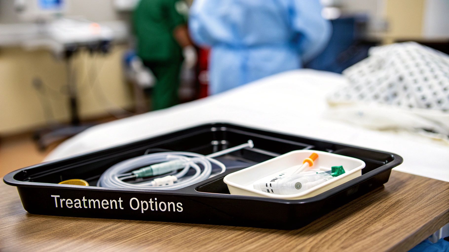
Once your electrophysiologist has that detailed electrical map of your heart and knows exactly where the trouble spot is, the conversation turns to solutions. Thankfully, we have an incredible range of treatments that can restore your heart’s steady rhythm and give you your life back. The goal is always the same: fix the underlying electrical fault so your heart can get back to doing its job.
The right path forward depends entirely on your specific diagnosis—what kind of arrhythmia you have, how often it happens, and how it’s impacting you. For some people, medication is the perfect starting point. For others, a more direct, hands-on procedure offers the best shot at a long-term fix. Let’s walk through the most common and effective treatments we use today.
Catheter Ablation: Correcting the Heart’s Short Circuits
For many fast arrhythmias like SVT and atrial fibrillation, catheter ablation is a game-changer. It’s a minimally invasive procedure that often provides a permanent cure. Think of it as a precision electrical repair job, all done from inside the heart. We often perform it right after a diagnostic EP study, using the very map we just created to guide the treatment.
During the procedure, the electrophysiologist threads a special catheter to the exact spot of heart tissue causing the short circuit. Then, we deliver a tiny, targeted burst of energy—either heat (radiofrequency) or cold (cryo)—to that tissue. This creates a tiny scar that acts as a roadblock, blocking the haywire electrical signals without harming the heart’s overall function. The abnormal pathway is gone, and the heart’s rhythm returns to normal. It’s become the go-to solution for many patients who want a real cure, not just years of managing symptoms with medication.
Implantable Devices: Pacemakers and ICDs
What if the problem isn’t a “short circuit” but a heart that’s beating too slowly, or one at risk of dangerously fast rhythms? That’s where implantable devices come in. These small but incredibly smart devices are placed just under the skin in the upper chest during a minor procedure, offering round-the-clock protection.
The technology behind these devices has a rich history that has completely transformed patient outcomes. The journey started back in 1952 with the first external pacemaker. By 1959, we had the first version that could be permanently implanted inside the body. Then, in 1970, the implantable cardioverter defibrillator (ICD) was invented to automatically stop life-threatening arrhythmias in their tracks. Today, millions of people around the world live full lives thanks to these advanced devices. You can read more about these therapeutic milestones and the evolution of cardiac rhythm devices.
These amazing devices fall into two main categories:
- Pacemakers: We use these to treat bradycardia, which is a slow heart rate. A pacemaker keeps a constant watch on your heart’s rhythm. If it senses your heart is beating too slowly, it sends a tiny, painless electrical signal to prompt a normal heartbeat. It’s like a safety net, making sure your heart rate never drops to a dangerous level.
- Implantable Cardioverter-Defibrillators (ICDs): An ICD is a more powerful device for patients at risk of life-threateningly fast ventricular arrhythmias. It can do everything a pacemaker can, but its most critical job is to detect a dangerously rapid rhythm and deliver a life-saving electrical shock to reset the heart. It acts as a constant guardian, protecting you from sudden cardiac arrest.
These devices are more than just treatments; they’re 24/7 monitors that provide incredible peace of mind. They work quietly in the background, allowing you to live your life with confidence, knowing you are protected.
The Role of Antiarrhythmic Medications
Before we jump to a procedure, and sometimes alongside one, medications play a vital role in managing heart rhythm disorders. Antiarrhythmic drugs work in different ways to help control your heart rate and rhythm. Some of these drugs slow down the electrical signals as they move through the heart, while others make the heart muscle itself less prone to firing off extra beats.
Medications can be very effective at controlling symptoms and making arrhythmia episodes happen less often. The key thing to know is that they typically manage the condition rather than cure it, which often means taking them long-term. Your electrophysiologist will walk you through the benefits and potential side effects to decide if medication is the right choice for your treatment plan. This is a crucial conversation, especially when considering all your options and finding the best hospital for heart surgery options.
Ultimately, the best treatment is the one that’s perfectly matched to your specific condition and your personal health goals.
Preparing for Your Electrophysiology Appointment
Taking an active role in your own healthcare is one of the most powerful things you can do. When you’re dealing with a potential heart rhythm issue, preparing for your consultation with an electrophysiologist can make all the difference. It helps you get the most out of your visit, turning what can be a nerve-wracking experience into a truly productive conversation.
The first step, of course, is choosing the right specialist. When you start to find a doctor, look for an EP who is board-certified in both cardiovascular disease and clinical cardiac electrophysiology. That dual certification is a strong signal that they have deep, specialized training in this complex field.
How to Get Ready for Your Visit
To have a truly effective appointment, the best thing you can do is walk in with your information organized. This gives your doctor a clear, immediate picture of your health and what you’ve been experiencing.
- Document Your Symptoms: Keep a simple journal of your symptoms. Note what the feeling is—a flutter, a racing heart, a skipped beat—and when it happens. Jot down how long it lasts and what you were doing right before it started. Every detail helps.
- Gather Your Medical History: Bring copies of any past heart-related tests you’ve had, like ECGs, stress tests, or Holter monitor reports. If you were referred by another physician, make sure their notes have been sent over ahead of your appointment.
- List All Medications: Make a complete list of everything you take. This means prescriptions, over-the-counter drugs, vitamins, and even herbal supplements. Don’t forget to include the dosage for each one.
Coming prepared means your electrophysiologist can spend less time piecing together your history and more time focused on you.
Essential Questions to Ask Your Doctor
Remember, your appointment is a conversation, not a lecture. Asking smart questions is the key to understanding your condition and the path forward. It’s a great idea to write your questions down ahead of time so you don’t forget anything in the moment.
Having your questions ready transforms you from a passive recipient of information into an active participant in your own care. It’s about building a partnership with your doctor based on clarity and shared goals.
Here are a few questions you might want to start with:
- From what you’ve heard so far, what do you think my specific diagnosis might be?
- What kind of tests do you recommend, and what will each one tell us about my heart?
- Could you walk me through all of my treatment options, from lifestyle changes and medication to procedures?
- For each of those options, what are the typical success rates and potential risks?
- What can I be doing at home—in terms of diet, exercise, or stress—to support my heart health right now?
Your Electrophysiology Questions, Answered
When you’re dealing with your heart health, it’s natural to have questions. This last section tackles some of the most common things patients ask about cardiac electrophysiology, giving you straightforward answers to build your confidence and understanding.
What Is the Difference Between a Cardiologist and an Electrophysiologist?
Think of it like this: a general cardiologist is your all-around heart doctor. They’re experts in the heart’s plumbing and structure, managing conditions like blocked arteries, valve problems, and heart failure.
A cardiac electrophysiologist, or EP, is a cardiologist who has gone on to complete 1-2 more years of highly specialized training. Their entire focus is on one thing: the heart’s electrical system. So, while your cardiologist might be the one to first spot an irregular heartbeat, they’ll likely send you to an EP for the high-tech diagnostics and specialized treatments like an ablation or a pacemaker.
Is an Electrophysiology Study Painful?
Most people find that an EP study isn’t painful. You’ll be given sedation to help you stay completely relaxed and comfortable, and the team will use a local anesthetic to numb the area where the catheters are inserted—usually in your groin.
You might feel a quick pinch from the numbing shot and a bit of pressure as the catheters are gently moved into place. Sometimes, the EP needs to briefly trigger your arrhythmia to see exactly where it’s coming from, so you might feel your familiar palpitations for a moment. But the team is right there with you, monitoring everything to make sure you’re okay.
The top priorities during an EP study are your safety and comfort. The sedation and local anesthetic work very well to keep any discomfort to a minimum while the team gets the vital information they need.
How Long Is Recovery After Catheter Ablation?
The good news is that recovery after a catheter ablation is usually very quick. Most people stay in the hospital overnight just so the team can keep a close eye on them.
When you get home, the main instruction is to take it easy for a few days. You’ll need to avoid any strenuous exercise or heavy lifting to let the small incision site heal up. Many patients are back to their normal, non-strenuous daily activities within about a week. It’s also normal to feel some minor chest soreness or even a few palpitations as your heart heals over the next few weeks. Your doctor will give you a clear, personalized recovery plan to follow.
Finding a specialist with the right expertise is a critical step in your healthcare journey. Haute MD offers a curated network of the nation’s leading, board-certified physicians, including top cardiac electrophysiologists. Explore our directory to connect with a trusted expert near you and take the next step toward better heart health. Find your specialist on Haute MD.